Structure and Function of parts of the human eye
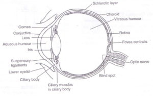
The mammalian eye is spherical, fluid-filled structure whose wall consists of 3 distinct layers:
- The outer layer or sclera (sclerotic layer)
- The middle layer or choroids
- The inner layer or retina
Sclera
Sclera is a white fibrous layer which protects the delicate inner parts of the eyeball and helps in maintaining its shape.
The sclera continues and forms the cornea at the front of the eye.
The cornea is a transparent layer which allows light to enter the eye. It also refracts the light entering the eye.
Covering the front portion at the cornea is a protective thin transparent membrane called conjunctiva. It is continuous with the epithelium of the eyelid.
Choroid
It is a dark pigmented, membranous middle layer that contains numerous blood vessels.
Its function is to absorb stray light to prevent internal reflection within the eye and to provide nourishment to the eye.
At the front of the eye, the choroid extends and forms the ciliary body and iris.
The iris is a thin round sheet of muscular tissue. It contains two sets of muscles, circular and radial muscles which control the diameter of the pupil.
The iris is pigmented giving the eye its colour e.g. black, brown or blue.
The pupil is the opening in the iris which allows light to enter the eye.
The ciliary body is an extension of the choroids, the iris and suspensory ligaments attached to it.
It contains circular and smooth muscles which contract and relax to alter the shape of the lens.
The ciliary body secretes the aqueous humour.
The lens is a transparent biconvex structure located immediately behind the pupil of the vertebrate eye.
It is held in position by the suspensory ligaments, which become taut or loose to alter the shape of the lens.
The lens divides the eyeball into anterior and posterior chambers.
The anterior chamber i.e. that part behind the cornea, is filled with a watery fluid the aqueous humour while the posterior chamber i.e. the part between the lens and the retina is filled with a denser, jelly-like transparent material called the vitreous humour.
Aqueous and vitreous humour maintain the spherical shape of the eyeball, and also refract incoming light towards the retina.
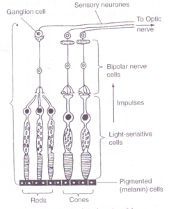
Retina
The retina is the light-sensitive layer composed of three regions;
- An outer pigmented region in contact with the choroid.
- A middle region of photoreceptors consisting of rods and cones.
- An innermost region of neurons.
The neurons run over the surface of the retina and join to form the optic nerve which transmits nerve impulses from the retina to the brain for interpretation.